
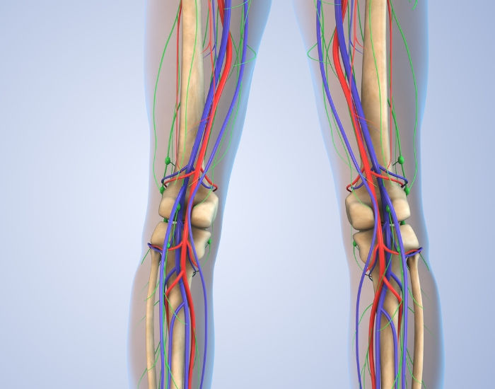
The genicular arteries are a network of arteries around the knee joint that contribute to the blood supply of the knee. Understanding these variations in the anatomy of genicular arteries is important in clinical practice, particularly in procedures such as angiography, arterial interventions, and surgical approaches to the knee joint. Knowledge of variant anatomy helps healthcare professionals anticipate potential challenges and complications and adapt their approach accordingly.

Meet your vein specialist
Dr. Syed Raza, MD
Vascular & Interventional Radiology · Founder, Premier Vein & Vascular Center
Fellowship-trained interventional radiologist certified by the American Board of Radiology and the Royal College of Radiologists (UK). Former Chief of Interventional Radiology at UTMB Galveston and Memorial Hermann Memorial City, with 15+ years treating varicose veins, venous reflux and peripheral arterial disease.
Arteries in Legs Anatomy and the Vascular Network Behind the Knee
The arteries in the legs anatomy features an intricate system responsible for supplying oxygen-rich blood to the lower limbs. One of the key components is the group of arteries behind the knee, primarily derived from the popliteal artery. These include the genicular branches, which form an essential network ensuring perfusion of the knee structures. Among these, the middle genicular artery holds particular importance—it enters the joint capsule and nourishes the synovial membrane and cruciate ligaments. Proper understanding of this vascular network is vital in planning interventions and preventing inadvertent injury during knee surgeries.
Quick Answer: The genicular arteries are a group of small arteries that encircle the knee and supply the joint lining, ligaments, bone and surrounding tissues. They usually include the descending genicular artery from the femoral artery, plus the superior medial and lateral, middle, and inferior medial and lateral genicular arteries from the popliteal artery, with recurrent branches of the tibial arteries joining the network. Their number, origin and course vary between people, which is why interventional radiologists map each patient’s arteries with angiography before procedures such as genicular artery embolization.

Origin and Branches of the Genicular Arteries
The genicular arteries arise from various branches of the femoral artery, popliteal artery, and the descending genicular artery.
- Branches: The genicular arteries have several branches that form an intricate network around the knee joint. These branches include the superior medial genicular artery, superior lateral genicular artery, middle genicular artery, inferior medial genicular artery, and inferior lateral genicular artery.
- Distribution: These arteries distribute blood to different regions of the knee joint. The superior medial and lateral genicular arteries supply blood to the proximal part of the knee joint, including the femur and surrounding tissues. The middle genicular artery supplies blood to the synovial membrane within the joint capsule. The inferior medial and lateral genicular arteries provide blood to the distal part of the knee joint, including the tibia and fibula.
- Anastomoses: The genicular arteries form numerous anastomoses (connections) with each other and with other arteries around the knee joint, such as the descending genicular artery, circumflex femoral arteries, and the anterior and posterior tibial arteries. These anastomoses create redundant pathways for blood flow, ensuring adequate perfusion of the knee joint even if one artery is compromised.
Variants of genicular arteries anatomy
Variants in the anatomy of genicular arteries are not uncommon and can include variations in their origin, course, branching pattern, and distribution. Here are some common variants:
- Additional branches: Variations can occur in the number and origin of branches of the genicular arteries. While the main branches typically include the superior medial, superior lateral, middle, inferior medial, and inferior lateral genicular arteries, additional branches or variations in their origins may be present.
- Absent or hypoplastic branches: In some individuals, one or more branches of the genicular arteries may be absent or significantly reduced in size (hypoplastic). This variation may not necessarily affect the overall blood supply to the knee joint but could influence the collateral circulation in cases of arterial occlusion or injury.
- High origin: Sometimes, the genicular arteries may have a higher origin than usual. For example, the superior medial genicular artery might arise directly from the femoral artery rather than from the popliteal artery.
- Low origin: Conversely, the genicular arteries may have a lower origin than usual. This could involve branches originating from arteries below the typical level, such as the descending genicular artery or the anterior tibial artery.
- Anomalous branches: Occasionally, anomalous branches or connections may be present, leading to variations in the arterial supply to specific regions of the knee joint. These anomalies may not follow the typical pattern observed in most individuals.
- Crossing branches: Some genicular arteries may exhibit crossing branches, where arteries from different sources intersect or overlap within the knee joint region. These crossings can create unique vascular patterns and potential points of vulnerability or collateral circulation.
- Variant pathways: In addition to variations in branching patterns, there can be differences in the pathways taken by the genicular arteries as they supply blood to different regions of the knee joint. These variant pathways may reflect adaptations to individual anatomy or developmental anomalies.
Why Understanding Genicular Artery Variants Matters
Understanding the arteries behind the knee is essential not only for diagnosing vascular conditions but also for planning surgeries and minimally invasive procedures. In treatments like genicular artery embolization (GAE) for knee pain or during trauma surgery, awareness of a patient’s specific genicular artery anatomy can mean the difference between success and complication. Accurate imaging and expertise are required to navigate this network safely.
Contact us today to learn more about the complex vascular system of the knee joint and how it may impact your overall health. Our team of experts at Premier Vein and Vascular Center specializes in diagnosing and treating vascular conditions, helping you maintain optimal blood flow and function in all areas of your body. Don’t let potential anomalies or variations in your genicular arteries go unnoticed – schedule a consultation with us today.
Genicular Arteries at a Glance
For patients reading about knee procedures, the names of these arteries can be hard to keep straight. The table below summarizes the usual pattern described in the anatomy above, along with why each vessel matters in practice. Keep in mind that the variants already discussed mean any one person may differ from this textbook picture.
| Artery | Usual origin | Main region supplied | Practical relevance |
|---|---|---|---|
| Descending genicular | Femoral artery, just above the knee | Inner side of the knee and lower thigh | Often involved when knee pain is on the inner side |
| Superior medial genicular | Popliteal artery, above the joint | Inner upper knee, lower femur | Commonly evaluated in medial knee pain |
| Superior lateral genicular | Popliteal artery, above the joint | Outer upper knee, lower femur | Commonly evaluated in lateral knee pain |
| Middle genicular | Popliteal artery, at joint level | Joint capsule, cruciate ligaments, synovium | Supplies deep structures inside the joint |
| Inferior medial genicular | Popliteal artery, below the joint | Inner lower knee, upper tibia | Often contributes to medial joint-line pain |
| Inferior lateral genicular | Popliteal artery, below the joint | Outer lower knee, around the fibular head | Runs close to the lateral meniscus |
| Recurrent tibial branches | Anterior and posterior tibial arteries | Front and lower knee | Can supply the area below the kneecap |
How the Genicular Arteries Work as a Network
The most useful way to picture the genicular arteries is not as separate pipes but as a ring of connected vessels around the knee, sometimes called the genicular anastomosis. Because the branches join one another above, below and around the kneecap, blood can reach most parts of the knee by more than one route.
This redundancy has two important consequences. First, it protects the knee. If one branch is narrowed or blocked, neighboring branches can usually keep the tissues supplied. Second, it explains why closing a few very small branches during a procedure does not usually harm the healthy structures of the knee: the main blood flow to the leg, through the popliteal artery and its continuation into the lower leg, is left untouched, and the network continues to nourish the joint.
The same network becomes very important in people with peripheral arterial disease. When the main thigh artery becomes blocked, the genicular branches often enlarge over months and years to act as natural bypasses, carrying blood around the blockage to the lower leg. On angiography these collateral vessels can be seen clearly, and they are one reason some people with significant arterial blockages still have reasonable circulation to the foot.
Why Genicular Artery Anatomy Matters for Knee Pain Treatment
In osteoarthritis, the inflamed lining of the knee develops many tiny new blood vessels that branch from the genicular arteries. These abnormal vessels, together with the nerve fibers that grow alongside them, are thought to contribute to chronic knee pain. Genicular artery embolization aims to reduce that abnormal blood flow by delivering microscopic particles into the small branches feeding the inflamed areas.
To do that precisely, the interventional radiologist must know which arteries supply the painful part of each patient’s knee. In general, pain on the inner side of the knee tends to involve the descending genicular and medial genicular branches, and pain on the outer side tends to involve the lateral branches. But because of the variants described above, such as a high origin, an absent branch or an extra vessel, the real pattern in any individual is confirmed only on imaging during the procedure.
How the arteries are mapped
- Angiography. Through a small puncture, a thin catheter is guided into the leg arteries and contrast dye is injected while X-ray images are taken. This shows each genicular branch, where it starts and where it goes, and highlights areas of abnormal vessel blush around the inflamed joint lining.
- Cone-beam CT. Some interventional radiologists use a rotating X-ray scan during the procedure to produce three-dimensional images, which help confirm exactly which tissue a branch supplies before treatment.
- Careful catheter positioning. Very small microcatheters are advanced into the chosen branches so that particles are delivered only where intended.
Protecting the skin and nearby structures
Some genicular branches also send small vessels to the skin over the knee. If particles reach those vessels, patients may notice temporary skin discoloration, which usually fades on its own. Knowing the anatomy and its variations, and checking each branch on imaging before treatment, helps reduce this and other unintended effects. Patients sometimes have ice applied to the skin over the knee during the procedure for the same reason.
How We Evaluate Knee Pain Before Considering GAE
Anatomy matters during the procedure, but the decision to treat starts with a careful evaluation. At Premier Vein & Vascular Center, Dr. Syed Raza, an interventional radiologist, follows these steps:
- Focused history. We ask exactly where the knee hurts, what makes it worse, how long it has lasted and which treatments you have already tried, including physical therapy, medications and injections.
- Pain mapping and examination. We ask you to point to the most painful areas, check for swelling, tenderness and joint stability, and examine the hip and the circulation in the leg, since hip disease or arterial narrowing can mimic knee pain.
- Imaging review. Weight-bearing knee X-rays show the degree of osteoarthritis, and MRI may be reviewed or ordered when another cause of pain needs to be excluded.
- Circulation check when needed. If pulses are weak or there are symptoms of arterial disease, an ankle-brachial index or arterial ultrasound may be done before any procedure.
- A clear plan. We explain whether GAE is likely to help, what the procedure involves and how the arteries will be mapped, and the possible risks and recovery.
Other Conditions Involving the Arteries Around the Knee
The arteries behind the knee are relevant to more than knee pain. Conditions that interventional radiologists and vascular specialists commonly evaluate in this area include:
- Narrowing or blockage of the popliteal artery from atherosclerosis, which can cause calf cramping with walking and may rely on genicular collaterals to keep the lower leg supplied.
- Popliteal artery aneurysm, a ballooning of the artery behind the knee that can form clots. It may be felt as a pulsing lump and is checked with ultrasound.
- Bleeding after knee surgery or injury, where a small genicular branch is damaged. These bleeds can sometimes be stopped by embolizing the injured branch through a catheter, avoiding further surgery.
- Trauma around the knee, such as dislocation, where the arteries need prompt assessment.
Call 911 or seek emergency care if you notice:
- A foot or lower leg that suddenly becomes cold, pale, blue or numb
- Sudden severe leg pain with weakness or inability to move the foot
- A rapidly growing, painful or pulsating swelling behind the knee
- Heavy bleeding or a growing lump at a catheter puncture site after a procedure
- A hot, red, very swollen knee with fever
When to See a Specialist in Houston
If you live with knee pain from osteoarthritis that has not improved with physical therapy, medication or injections, or if you have questions about the circulation around your knee, an interventional radiology opinion can help. Dr. Syed Raza is a fellowship-trained interventional radiologist and former Chief of Interventional Radiology at UTMB Galveston and Memorial Hermann Memorial City. Our team at Premier Vein & Vascular Center in Houston reviews your imaging, explains your options in plain terms and, when appropriate, performs GAE using careful arterial mapping. You can learn more on our knee pain treatment page. Most insurance plans, including Medicare, are accepted, and our staff can check coverage for you.
Talk With a Specialist at Premier Vein
Have questions about your symptoms or treatment options? Dr. Syed Raza and the team at Premier Vein & Vascular Center can evaluate your concerns and explain your options clearly. Same-week appointments are usually available, and most insurance plans are accepted.
Frequently Asked Questions
How many genicular arteries are there?
Most descriptions include five main genicular arteries from the popliteal artery (superior medial, superior lateral, middle, inferior medial and inferior lateral) plus the descending genicular artery from the femoral artery. Recurrent branches of the tibial arteries also join the network, and the exact number varies between people.
Is it safe to block genicular arteries during GAE?
GAE targets only very small branches feeding inflamed tissue, while the main artery to the lower leg is left open. Because the genicular arteries form a connected network, the knee continues to receive blood through other routes. As with any procedure, there are risks, which your doctor will discuss.
Why do genicular arteries vary between people?
Blood vessels form in many small steps during development, and slight differences in which vessels persist lead to variations in origin, size and branching. These variants are usually harmless but are important to recognize during procedures.
Can you see the genicular arteries on an MRI?
Standard knee MRI is designed to show bone, cartilage and soft tissue and does not reliably show these small arteries. Angiography during a procedure is the most detailed way to see them, and CT or MR angiography can be used in some situations.
Do the genicular arteries help if the main leg artery is blocked?
Yes. When the femoral or popliteal artery becomes narrowed or blocked, the genicular arteries can enlarge over time and act as natural bypass routes. This collateral flow helps keep blood reaching the lower leg and foot.
ABOUT PREMIER VEIN & VASCULAR CENTER: Premier Vein & Vascular Center, a leading vein clinic in Houston and among the top specialized vein treatment clinics in the USA, has provided expert vein care for over 20 years. The center offers minimally invasive treatments for varicose veins, spider veins, and deep vein thrombosis, led by Dr. Syed Raza, MD, a Board-Certified Vein Specialist and Interventional Radiologist. Dedicated to pain-free, healthier living, the clinic delivers cutting-edge, patient-centered care for optimal results.
Trusted Health Resources
- Osteoarthritis, National Institute of Arthritis and Musculoskeletal and Skin Diseases (NIH)
- Osteoarthritis, Centers for Disease Control and Prevention