Breast Lump/Mass Biopsy in Houston, TX
Breast biopsy is performed when a breast lump is detected by physical examination, ultrasound, or mammography. The terms, “lump” and “mass” are used interchangeably in medical terminology.
A diagnostic report is generated after seeing the breast mass with these imaging modalities. The description of breast mass from the diagnostic report usually places the mass in one of the four quadrants of breast, upper outer, upper inner, lower outer and lower inner quadrants. Using a clock, further refines the position by assigning a number to position of mass, such as 11, o clock or 7, o clock from nipple etc. Typical description also includes the distance of breast mass from the nipple and its depth from skin surface.
If the breast mass is visible on ultrasound, a biopsy can be performed using ultrasound guidance. Breast biopsy is performed in the office, and it is relatively simple and quick. After placing the patient in a comfortable supine position on the table, skin of breast is cleaned with an antibacterial solution in a sterile fashion. Using ultrasound guidance to localize the mass, local anesthesia is infiltrated in the skin and a needle is advanced into breast mass under direct vision, with ultrasound showing the exact needle trajectory into the mass (Figure 1). Multiple samples are then taken from the mass, using the biopsy needle, and placed in Formalin solution. Patients are typically able to go home immediately after biopsy if procedure has been performed under local anesthesia. The samples collected during biopsy are dispatched to pathologist for examination under microscope and generating a histopathology report for oncologist which may take 2-4 working days.
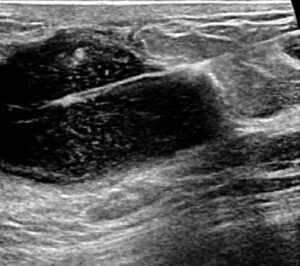
Figure 1, dark area on ultrasound picture is the breast mass and bright white line is the needle taking sample from breast mass.